Introduction
The Ultrasound Overlay Display is an "Editor" in MITK-speak, meaning that it fills the centre part of the screen, and provides a view of the data in the Data Manager. It is a custom viewer for providing augmented reality displays, merging rendered surface meshes and ultrasound data.
Usage
Imagine you have an ultrasound machine providing ultrasound images, connected via the Data Sources View (see Figure 1).
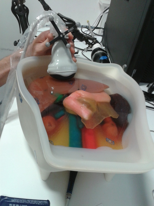
Figure 1: Physical layout of a tracked (NDI Vicra) ultrasound probe, viewing an abdomen phantom.
Then the Ultrasound Overlay Display can be used to provide a Freehand Ultrasound system (see Figure 2).
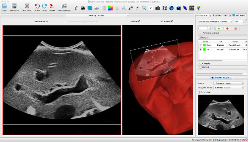
Figure 2: The Overlay Display merges ultrasound data with virtual rendered data (left) and displays the 3D imaging plane, moving in the 3D world (right).
The controls for the display itself are simple. See Figure 3.

Figure 3: The Controls.
- A combo box, labelled "please select" enables the user to select the image to track. Essentially the viewer orientates the left hand camera to always face this image.
In order to actually move the image according to some tracking data, you should use the Tracked Image plugin. This editor is JUST a viewer.
Preferences
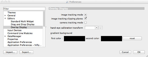
Figure 4: The user preferences.
Here we can see:
- "clipping planes" check box. If checked will place a clipping plane +/- 1mm from the image plane, creating a crude contouring effect, displaying the outline of surface meshes on the image data.